BetaBASE

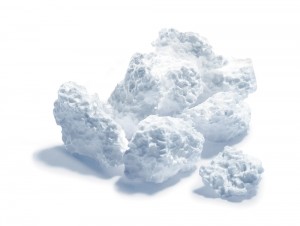
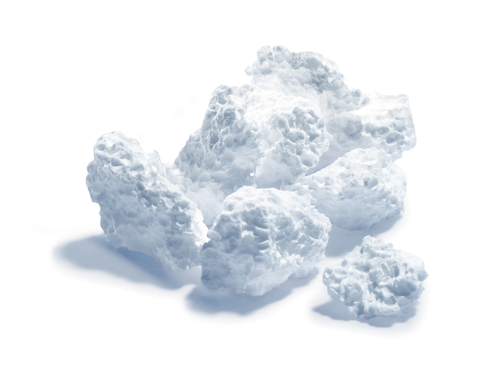
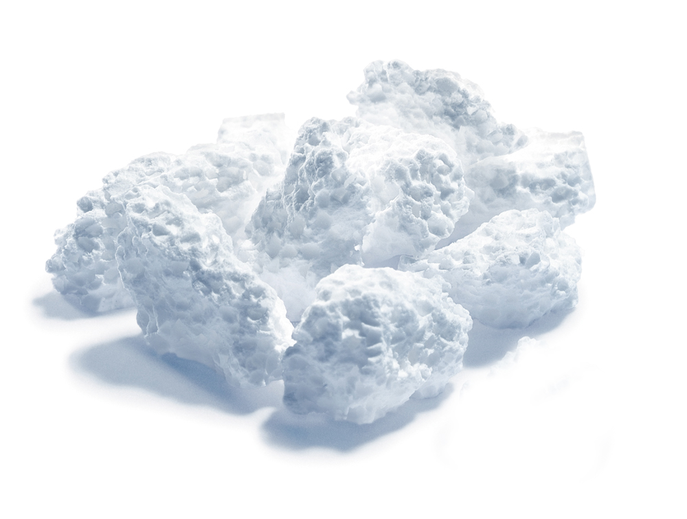
BetaBASE is a bioresorbable bone replacement from microporous and macroporous β-tricalcium phosphate. BetaBASE consists of pure-phase β-TCP.
Potential risks of infection involved in the use or processing of biological material (HIV, BSE, inter alia) are non-existent with BetaBASE.
BetaBASE has an interconnected pore system with micropores and macropores that reproduce the well-known osteoconductive effect almost perfectly.
Osteoblasts and blood vessels are able to proliferate rapidly in the open pore system and grow swiftly in the BetaBASE. This also enables internal resorption of the materials.
The high overall porosity of BetaBASE (> 60 %) means that the body has to break down a far smaller quantity of bone replacement material, based on the volume of the defect. This accelerates the resorption process and also creates new opportunities to fill large bone defects. Clinical studies have demonstrated that the material is completely resorbable after 9 to 12 months.
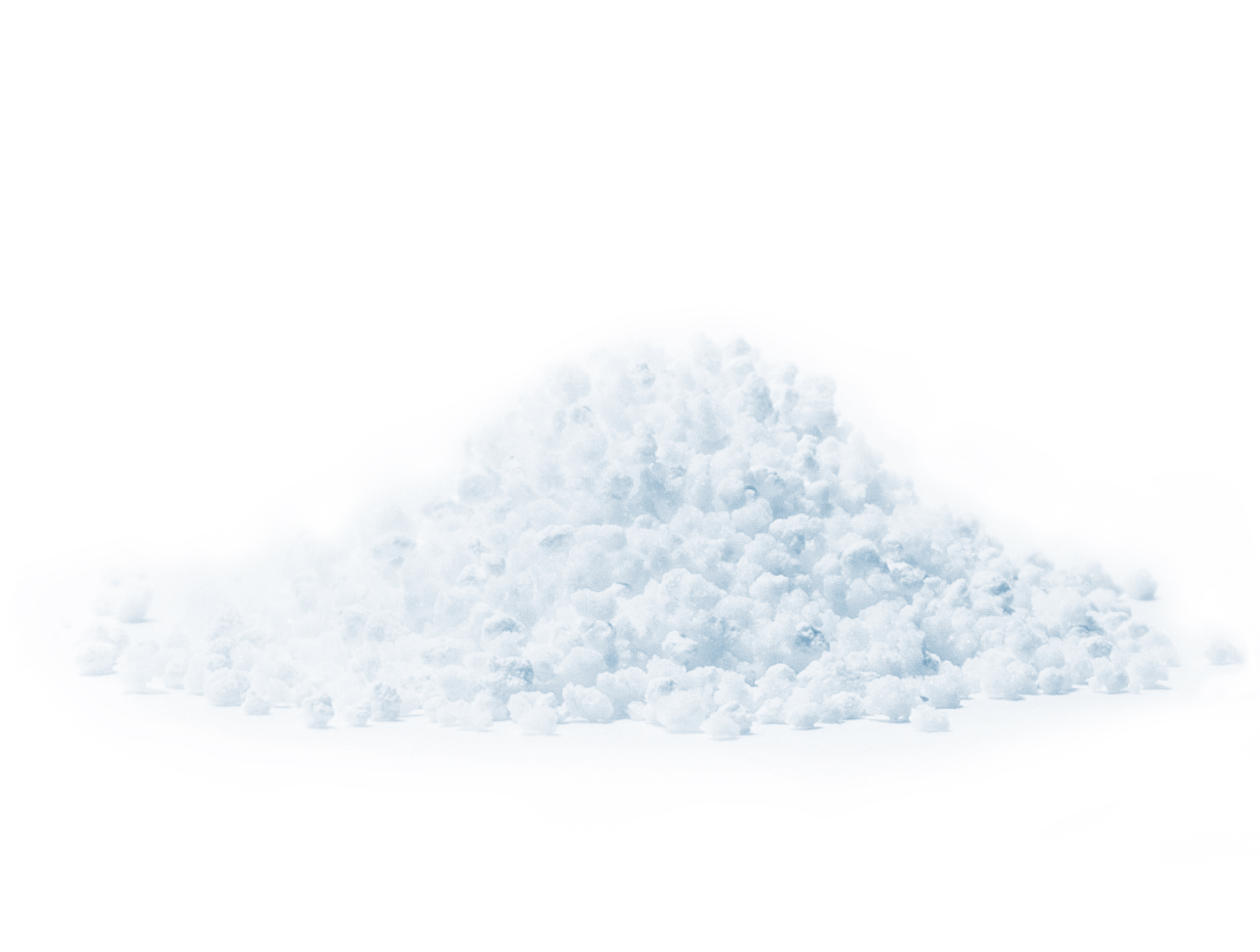
The polyhedric granular structure of Betabase makes it safe and easy to apply to the defect, in particular when mixed with the patient’s blood.
BetaBASE conforms to ASTM F 1088-04, the internationally recognised standard for materials.

Indications
Addition of the material to autogenic or allogenic spongiosa to reconstruct bone defects e.g. in cases of spondylodesis, vertebral body replacement and in joint replacement surgery.
- Filling defects in correction osteotomies
- Filling the sites from which autogenic bones have been removed
- Filling bone cysts
- Use in arthrodesis
- Filling defects after the removal of benign bone tumours
 Figure 1: Macropores up to 500 μm are visible.
 Figure 2: Micropores in detail, approx. 5 μm, sinter necks are visible.
Application
It is advisable to apply Betabase in a moist state. The patient‘s own blood or blood plasma should be used. If neither option is available in sufficient quantities, a sterile isotonic saline solution may be used.
Where defects are larger than approx. 2 cm³ it is advisable to mix Betabase with spongiosa.
Local administration of antibiotics is possible.
 Figure 3: Cyst filled with 30 ml.
Figure 4: The material is virtually invisible 16 months later.
 Figure 5: Mesh graft after correction osteotomy.
Figure 6: Good resorption after 10 months.
BetaBASE

BetaBASE MP
BioBASE
BioBASE AP
PolyPIN
EpiGARD